We understand that travelling to a specialist international radiotherapy clinic in another country is a huge decision to take, and when it is considered to be part of your cancer treatment plan, it is one of a series of significant and sometimes stressful choices to make.
Because of this we ensure that your stay in the beautiful city of Vienna is as comfortable, relaxing and stress-free as possible, with arrangements for extended stays, specialist rooms for people undergoing treatment and their loved ones, assistance with visa and medical documents and help with any other part of your stay in Austria.
We do this because of our multidisciplinary holistic focus, ensuring that every single part of your itinerary is focused on ensuring you are in the best possible condition for treatment and recovery, and we have considerable experience in giving international patients the best experience possible.
However, seeking treatment abroad is a huge decision and not one that is ever to be taken lightly, but here are some of the reasons why you may be considering or might have been recommended care at an international clinic.
You Need Access To Advanced Medical Equipment
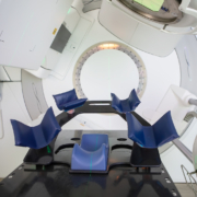
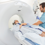
Radiotherapy is an incredible non-surgical non-invasive form of treatment that can be used to incredible results, but the cost of the equipment and the specialist expertise required to operate it safely and plan treatments around it means that not every nearby hospital has access to it.
This is particularly true for TTFields therapy, branded as Optune by the company NovoCure. As it is a state-of-the-art treatment, very few clinics in the world can provide it at all, so the alternative is to travel to a country where it is available.
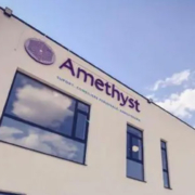
Amethyst has a wide range of radiotherapy clinics across Europe that have specialist medical equipment and world-class medical professionals that can treat specific conditions with equipment potentially unavailable in your country of origin.
Skip Waiting Lists
Alternatively, some treatments such as radiotherapy or chemotherapy are commonly available but the specific type of treatment you need for a particular cancer diagnosis may involve additional costs or having to go onto a waiting list to gain access to limited facilities.
If those facilities are available elsewhere and they are suitable for your treatment plan, then the best advice may be to travel to an international clinic and get treated sooner, particularly if there is a risk of progression if you need to wait.
At the same time, this does not mean rushing pre-treatment consultations, tests and scans, but instead ensuring that treatment can start as soon as you are ready so it can be completed and you can begin your recovery.
If You Are Able To
Your local doctor will be able to advise you if you can safely travel given the stage or extent of your cancer, and provide either medication or advice for how to take care of yourself during your journey and throughout the duration of your stay in another country.
We will be able to provide some advice as well because our focus is ensuring you get the best possible care in order to have the most positive outcome of any treatment.
Similarly, if you plan on flying to Vienna for treatment, then you may need to prove that you are fit to fly, in case your existing treatment plan, medications or any previous procedures could affect your health whilst in the air.
Depending on where you live, flying may not be necessary, and there are often alternative forms of transport such as long-distance rail or road if you need to travel but cannot fly.
Therapeutic Restorative Relaxation
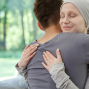
Travelling to another country for treatment should never be confused with a holiday, as the focus is consistently on your health condition. It is important to plan your itinerary around your recovery, rather than sightseeing. Once you recover, you can always come back.
However, you may get some of the mental and physical health benefits of a holiday by travelling to a beautiful new country and you may find clinical recovery outcomes are supported through stress reduction and better rest, both of which can help improve the body’s healing processes.