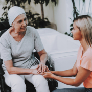
Because radiotherapy is a powerful, long-term treatment, its use is typically targeted carefully to ensure that its benefits are maximised and are proportionate to the significance and severity of the condition they are intended to treat.
This means that radiotherapy is primarily used to treat cancer, but it can sometimes be used to treat non-malignant conditions when it is suitable to do so, and there is a proven evidence base that it can be used to significantly help.
One particularly interesting example of how radiotherapy can be used to help treat conditions other than cancer is heterotopic ossification, one of several non-malignant conditions alongside osteoarthritis, chronic pain and Ledderhose disease that radiotherapy can help treat.
How is this the case? What causes heterotopic ossification in the first case? Can radiotherapy always be used? And what are the alternatives if it cannot?
What Is Heterotopic Ossification?
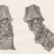
An inflammatory disease that can cause serious pain and decreased range of motion, heterotopic ossification (HO) is the growth of bone where it should not be, almost always replacing soft tissue or muscles.
It is sometimes mistaken for bone spurs, fragments or chips, in part because both HO most commonly emerge as a result of a traumatic injury, a response to surgery or have no cause at all.
It can vary in severity from a barely noticeable lump to a significant growth that restricts your range of motion, often requiring surgery to remove.
What is known in all cases, however, is that anyone who has experienced HO bone growth is at risk of it happening again, meaning that treatment is needed not only to remove the bone but also to prevent it from regrowing.
What Causes Heterotopic Ossification?
There are two different types of heterotypic ossification, which vary not only in root cause but also in severity.
The most common is nongenetic heterotopic ossification, which is caused by a traumatic injury that your body rebuilds too effectively.
They often occur following an accident which affects your joints, your head, your neck, your pelvis or anywhere else you are most likely to be injured, but they can also emerge as a response to the trauma inherent to surgery.
Sometimes it can grow for no reason at all, which can sometimes cause concern as the ossification is confused for cancer growth before it is tested and is ultimately benign.
Bone growth due to injury or surgery is not uncommon, happening in a third of people who have hip replacement surgery or treatment for a major tibia or fibula fracture in the leg. Most of these will not be noticeable, however.
Alternatively, HO can also be genetic in origin, most notably found with the very rare disorder fibrodysplasia ossificans progressiva (FOP), which causes bones to progressively form in connective tissue whenever it flares up.
Can Radiotherapy Always Help To Treat Heterotopic Ossification?
If bone growth caused by heterotopic ossification is causing problems, there are treatments that can help to cure it or prevent further bone growth, discomfort and pain.
Radiotherapy is not required for every case of heterotopic ossification. Many mild cases can be managed conservatively, and some areas of heterotopic ossification cause few or no symptoms.
Radiotherapy is most commonly considered for patients at higher risk of recurrence, particularly following orthopaedic surgery such as hip replacement or previous surgical removal of heterotopic bone.
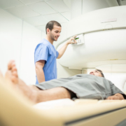
It is primarily used as a preventive treatment, particularly before surgery. A single dose of radiation is applied on the morning of the operation to stop the bone from growing in soft tissue and potentially causing damage to joint mobility.
It can also be used in the days following surgery, where the goal is to minimise the growth of bone in the wrong parts of your body.
As a result, it is typically used as an adjunctive therapy in combination with other treatments to ensure the best possible recovery.
What Treatments Are Used To Treat Heterotopic Ossification?
Alongside other preventative measures such as physiotherapy, bisphosphonates or ibuprofen, the main treatment to remove heterotopic ossification entirely is surgery.
As there is a risk that this surgery can also cause HO to form again, it is typically only used in cases where the bone growth has caused chronic pain or significant restrictions in movement, with radiotherapy used to reduce the chance of bone regrowth.
You typically need to wait until the bone has completely grown before it can be surgically removed, otherwise it will simply grow back and require further surgery.
This is particularly the case with genetically caused heterotopic ossification, such as that caused by FOP.
Radiotherapy can help to minimise bone growth by stopping bone cells from growing and dividing. As it is a very quick treatment that is accessible at many hospitals or nearby clinics, it has become best practice in order to assist with mobility.