The Die Presse Front Page

The team here at Amethyst Radiotherapy have been featured on the website of Austrian broadsheet newspaper Die Presse, showcasing the leading private radiotherapy clinic, based in Vienna, which provides a wide range of different radiation oncology services.

Centre director Paul Stuchetz spoke to the news source, explaining how Amethyst helps patients achieve the best possible courses of treatment to tackle their illness and detailing how the team works tirelessly to support people throughout their journey, with integrative cancer treatment at the heart of all they do.

Further information on radiation as a treatment option is also provided, with in-depth explanations as to how the external beam radiation device can be used to treat a range of different cancers, including prostate cancer, where it has seen particular success.

Linear accelerator (or Linac) radiotherapy uses high-energy X-rays and speeds up electrons to conform them to tumour shapes and sizes. When aimed at cancers, invasive cells are destroyed, but healthy ones go untargeted.

To find out more about such treatments, get in touch with the Amethyst Radiotherapy team today.

radiotherapy centre - Medical CT

There are certain conditions which can be effectively managed and treated once their causes are understood, and trigeminal neuralgia is a particularly illustrative example of this.

Once it has been diagnosed and a patient has been referred to a radiotherapy centre for treatment, several pathways are available determined by a patient’s medical history and the cause of pain.

As understanding is important to the treatment and management of the condition, here are some facts about trigeminal neuralgia that are important to know.

A Dentist Will Often Notice Symptoms First

The facial pain caused by trigeminal neuralgia is often felt in the gums, jaw and teeth, which usually means that it will be a dentist who will be the first person that many patients see when seeking treatment for the condition.

They can rule out that it is a toothache, abscess, or other dental concern through the use of an X-ray or dental CT scan, before suggesting that the patient sees their GP, who will themselves help to rule out other conditions that could cause similar pain sensations.

Once other causes are ruled out, a GP will refer the patient to a specialist or the patient will seek out a second opinion.

It Was First Discovered Three Centuries Ago

Trigeminal neuralgia is sometimes known as Fothergill disease, named after the doctor who first discovered the condition, John Fothergill.

In 1773, Dr Fothergill provided the first complete and accurate description of the condition, and would also describe conditions such as angina, diphtheria and streptococcal sore throat in English for the first time.

Nearly a century later, another doctor for the University of Edinburgh, John Murray Cornochan, would be the first person to successfully treat trigeminal neuralgia, through a surgical procedure to remove the trigeminal nerve.

There Are Three Main Types Of Trigeminal Neuralgia

Trigeminal neuralgia cases are grouped into three categories depending on the cause.

The first, and most common of these, is known as classical trigeminal neuralgia, which is caused by pressure on the trigeminal nerve which makes it activate and causes the facial pain that is characteristic of the disorder.

If the trigeminal neuralgia symptoms are caused by another medical condition, such as a tumour, multiple sclerosis or injury to the face, it is categorised as secondary trigeminal neuralgia.

Finally, if the cause is unknown, it is categorised as idiopathic trigeminal neuralgia, and until a cause is determined, treatment is centred around pain management.

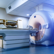
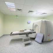
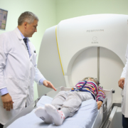
MRI Scans Can Find The Cause

In most cases, a diagnosis is confirmed through a magnetic resource imaging (MRI) scan, which uses a series of magnetic fields to create a detailed internal image of the face.

It can be used to find pressure on the trigeminal nerve, its location and the precise cause. This can be used to diagnose a patient’s symptoms as trigeminal neuralgia and is typically used to help plan treatment.

It Does Not Always Require Surgery To Treat

There are three main treatment paths, depending on the cause and how well a patient has responded to other treatments.

Initially, anticonvulsant medication is the first treatment many people with trigeminal neuralgia are likely to take.

Many over-the-counter painkillers are not effective at treating the specific cause of pain seen with trigeminal neuralgia, so regular doses of an anticonvulsant medication such as carbamazepine will be initially taken to slow down nerve impulses and stop the trigeminal nerve from activating.

Alternative medicines are available if carbamazepine does not work, but specialist surgery can also be offered which can provide relief for months, and sometimes years.

This includes microvascular decompression, which helps relieve pressure on the trigeminal nerve and can therefore provide long-term pain relief.

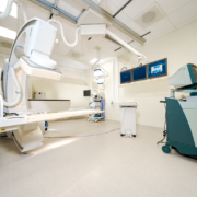
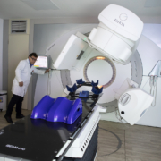
These also include keyhole surgical treatments undertaken under general anaesthetic which aim to deactivate the nerve entirely, but an alternative to this that specialists are using to treat trigeminal neuralgia without the need for surgery is stereotactic radiosurgery.

Treatments such as Gamma Knife work by using multiple beams of radiation concentrated to a point to provide precise doses of radiation to damage the trigeminal nerve where it enters the brainstem.

It works through the use of a complex frame which holds your head in place and is used to guide the beams of radiation to the central point where the nerve needs to be damaged in order to stop activating.

It requires no incision, no general anaesthetic (although local anaesthetic is often provided for the points where the frame is secured), and does not require a stay in the hospital once the procedure is completed.

It can sometimes take time for the procedure to take effect, but it can provide relief for years.

radiotherapy centre - Body scanner equipment in oncology department

Anyone who is prescribed a course of radiotherapy treatments from a specialist centre can feel comforted that they are receiving a proven treatment that is over a century old and is still evolving and improving in its efficacy.

From the very start of radiotherapy’s development as a potential treatment for cancer, the effectiveness of radiation was never in doubt, because the underlying therapeutic principle largely predates the discovery of radiation itself.

Replacing Chemicals And Electricity

The therapeutic action of radiotherapy is to burn away the tissue that is exposed to radiation, which means that its targeted use can effectively destroy unwanted cells.

The main developments from this discovery in 1896 by Leopold Freund, Eduard Schiff, Emil Grubbe and Victor Despeignes paved the way for progressively better targeted, safer and more effective forms of treatment.

However, the underlying therapeutic action was much older than this, even if the tools used to destroy the malignant tissue were far less accurate and far more dangerous.

Early 1800s: Electrotherapy

One main form of treatment which inspired the development of radiotherapy once radiation was discovered was electrotherapy.

Unlike modern electrotherapy, which tends to use small electrical currents to stimulate recovery or enhance the effects of certain chemotherapy treatments, the field of electrotherapy up until the early 19th century was rather more experimental.

Up until Golding Bird, who helped rehabilitate the field of electrotherapy in the same way that Henri Coutard would radiotherapy in the 1930s, electrotherapy was another treatment that was often extremely dangerous with somewhat dubious benefits.

It was used to stimulate skin tissue and potentially burn away unwanted cancerous cells, and this method of action still sees some use today in the treatment of varicose veins.

Early 1900s: Escharotics

One of the earliest forms of treatment for killing unwanted tissue was poisonous and caustic substances called escharotics. However, these substances are not only extremely dangerous, scarring and in some cases outright mutilating, they can cause permanent, potentially life-threatening damage.

They were popular in early medical practice up until the early 1900s because, without the knowledge of how tumours survive, the tumour did appear to be burned away, along with a significant chunk of healthy tissue, making it appear to be effective even if the tumour very often grew back.

Escharotics have been largely discredited by mainstream medical practice since, and many of the substances used are illegal to sell because of the significant amounts of damage they can cause.

Even the few forms of cancer that can be treated topically such as basal cell carcinoma are typically treated with less damaging and more effective topical solutions than caustic chemical burns.

1896: Discovery of radiation

As early as 1896, doctors knew that radiation could provide the same ablative effect, even if they did not quite understand why.

Victor Despeignes accidentally discovered the therapeutic effects of radiotherapy when trying to treat a patient with late-stage stomach cancer.

Whilst he ultimately did not survive and Mr Despeignes only opted for radiotherapy under the mistaken belief that cancer was caused by parasites, it inadvertently proved a principle that would be more effectively demonstrated by Mr Freund and Mr Schiff, and mark a seismic shift in the history of medicine.

1933: Stereotactic frame & electrotherapy

Interestingly, the first use of the stereotactic frame to treat a human patient involved electrotherapy.

In 1933, Martin Kirschner published a paper wherein he detailed his use of a treatment for trigeminal neuralgia by using a stereotactic frame to make an incision large enough to feed an electrode to the trigeminal nerve, burning it away and reducing the pain.

1940s: Gamma Knife development

This principle would later be used by Lars Leksell for his innovative Gamma Knife radiosurgery and serves to highlight just how influential pre-radiation radiotherapy treatments were and how similar their core method of action is.

The main difference, of course, is a matter of scale, effect and invasiveness to a patient.

Surgery was and remains very effective but is also extremely invasive and typically requires a patient to spend several nights in the hospital to recover.

Escharotics may not require incisions but they are invasive and require time to recover from the intense and often unpredictable damage that the materials could cause. This lack of consistency is what led to them being almost universally outlawed.

Electrotherapy was less invasive but in some cases still required an actual incision to provide the same effect of ablation and burning away of unwanted or harmful tissue.

2025: AI in radiotherapy

In early January 2025, researchers explored the potential of using AI to make what had become an already accurate set of treatments even more precise, something that has some benefits for brain lesions and tumours but can be even more important for treating cancers that affect the digestive and reproductive systems.

Prostate cancer is a disease that kills millions of men every year. It is one of the most common forms of cancer and men will increasingly become vulnerable to it after the age of 50. Those who are overweight, have a family history of the disease or who belong to particular ethnic groups are at higher risk, with black men facing the greatest peril.

Even without being in a high-risk group, men should get themselves checked out if they suspect something is wrong, due to issues like erectile dysfunction, urinary problems, blood in semen or unexpected weight loss.

Improving Diagnostics

As with all cancers, early diagnosis increases the chances of beating the disease. Indeed, there has been some good news on this front, with the UK-based University of East Anglia claiming a breakthrough in developing a test to show if the prostate cancer is benign (as the majority are) or aggressive, something the current blood test cannot do.

It works through a urine testing kit that can be delivered to homes and taken there, before being posted back to the lab. The contents of the test can now reveal a wide range of results, including clues that show that the patient should go for further testing.

Professor Colin Cooper, who headed up the team that developed the test, remarked: “It’s astonishingly exciting – this is potentially a game-changer both for the initial assessment for prostate cancer, and people who have been diagnosed.”

What this highlights is the reality that diagnostics have been a weak point in prostate cancer treatment up until now, which means fewer men get the diagnosis they need until the cancer is more advanced.

In some cases, that means the treatment and the path to beating the disease is longer and more gruelling than it might otherwise have been, while in others the best that can be achieved is to extend life rather than save it.

Radiotherapy And Other Treatments

Our radiotherapy centre treats many patients with the condition, helping fight the cancer effectively. However, the success of this may depend on how early diagnosis takes place.

The good news is that it is not just the ability to diagnose aggressive cancers that is improving. The range of treatments and the capacity to apply them in the most effective way has grown markedly in recent years. As an article in Indian website News X notes, there have been several major developments in recent years aiding survival rates.

Some of this includes other treatments that can complement radiotherapy, such as PARP inhibitors, which block the enzyme that helps cancer cells repair their DNA after it has been damaged by radiotherapy or other treatments. This can be critical in the success of radiotherapy, with advancements in this area proving invaluable.

Others work in their own way, such as hormone therapy, which has been around for many years and helps reduce the production of hormones that promote the growth of cancerous cells. In recent years new therapies have become more effective at this.

Immunotherapies also have a role to play, but some of the most exciting developments come in areas that enhance the effectiveness of radiotherapy in destroying cancerous cells. This includes radiogland therapy, which specifically targets PMSA a protein found on the surface of prostate cells, enabling the radiation to be aimed directly at tumours.

All this can be added to the effectiveness of improved scanning technology for early diagnosis. The University of East Anglia breakthrough could be a further step forward in this crucial area, but already there are other promising advancements, like the use of artificial intelligence to identify problems a radiographer might not be able to spot so easily.

Could Vaccines Offer A Future Hope?

Some have even talked about the potential of vaccines to help tackle the disease. Various possible kinds could achieve effective results in preventing prostate cancer from taking hold in the first place: peptide, nucleic acid, viral, cellular and dendric cell vaccines are all the subject of research.

Should any of these prove truly effective, it may be that one day the way prostate cancer is treated can be completely different, with a greater focus on prevention in much the way that the HPV vaccine can protect women against cervical cancer.

However, unless and until such vaccines are fully developed and proven to be effective, other treatments will still be needed for patients who have prostate cancer and are either seeking to beat the disease outright or at least extend their lives.

Radiotherapy will remain a crucial weapon in the armoury for the foreseeable future, working alongside an array of other treatments.

radiotherapy centre - Gamma Knife

If someone is prescribed a treatment plan that consists of stereotactic radiosurgery, they can enter the radiotherapy centre confident that they are being treated by one of the most effective, accurate and oldest radiosurgical treatments available.

The principle of stereotactic radiosurgery, as pioneered by Lars Leksell’s Gamma Knife in the late 1940s, is that multiple small beams of radiation that converge on a particular point can have a much greater effect without damaging the surrounding tissue and minimising overall exposure.

This treatment, whilst adjusted and refined over the past half-century, has been consistently used and effective even before there were effective ways to map treatments using MRI and CT scans. 

This naturally leads to a big question; if the Gamma Knife is so effective, why has the stereotactic principle not been widely applied to other forms of radiotherapy treatment outside of the brain and spine?

The answer is complex and one that medical researchers are doing their utmost to try and change.

The Sea Of Life

The almost tautological answer for why the Gamma Knife specifically is not used to treat other parts of the body is that it was only designed for the brain and relies on a very specific set of medical apparatus that has only been used in the head and cannot be used anywhere else.

Part of this is that the frame system that keeps the head in place for a Gamma Knife treatment is based on the Horsley-Clarke frame initially used to create an atlas of various animal brains before being applied to the human head over the course of four decades.

This tool, albeit heavily modified by Mr Leksell and others, is as fundamental and vital to the success of Gamma Knife as the source of radiation itself. This is why the treatment was tested and used before the widespread availability of three-dimensional medical imaging.

It meant that thanks to some rather complex mapping, a series of skull X-rays could be triangulated to position the stereotactic beams in order to treat patients effectively. Once CT scans and MRIs came in, this only made the process even more accurate.

A lot of this can be credited to Mr Leksell’s work himself, who famously said there was no tool too accurate for use on the brain, but whilst an accurate approach is mandatory for obvious reasons, it is also, relatively speaking, easier to achieve.

The skull helps keep everything in place and there is less variation, relatively speaking, with tumour and lesion positions, allowing for consistency and greater accuracy. 

Pair this with the stereotactic frame keeping the head from moving during treatments and it is perhaps understandable how the Gamma Knife started as a highly effective targeted treatment and has only gotten more accurate with time.

By contrast, the rest of the body is far less consistent and mobile. Organs are constantly moving in the body, not just in terms of pulsing, contracting and relaxing, but also shifting positions based on the position of the body.

Organs change shape depending on their use, muscles such as the diaphragm affect their shape, and even posture and the way people lie down can alter the shape, size and position of organs, particularly those in the abdomen.

This is a major problem when planning radiotherapy because the act of breathing creates a moving target that affects treatments, often requiring wider beams and more collateral damage to healthy tissue to ensure that all cancerous cells are destroyed.

There are ways to mitigate some of these issues, such as through the use of frames and casts to at least keep a body part securely in place during treatment, but there are a lot more variations than there are for the brain, requiring not only different treatments but a different treatment philosophy.

There have been attempts to change this over the years, primarily through increasing the speed at which medical images are produced and interpreted, and this could lead to Gamma Knife-like treatments becoming possible with that level of accuracy in the future.

In order for this to be accomplished, however, there needs to be a close to real-time form of three-dimensional imaging of body parts that can be used to plan treatments the same day they are executed, as unlike the brain there is the potential for much greater organ movement.

This concept, known broadly as real-time adaptive radiotherapy, is one that is at least conceptually possible, but it requires a widely-used real-time imaging system that, as of 2024 at least, has not been widely used.

doctors prepares the patient for the procedure on the Gamma Knife

Precision is key when it comes to any treatment related to the brain, and part of the reason why people are often prescribed a trip to a radiotherapy centre for stereotactic radiosurgery is precisely because of this precision.

Lars Leksell once noted that no tool can be too refined for the human brain, and this became the motivation for him, alongside a desire for bloodless neurosurgery, to develop what became the Gamma Knife, the first-ever stereotactic radiosurgery technique and still one of the most widely used in the world.

It is a critical piece of equipment that has helped to ease the suffering of countless people and help treat many different types of brain cancer. However, who was the first to receive treatment using this pioneering technique? 

It depends somewhat on the definition of stereotactic radiosurgery.

Stereotactic Surgery With Radiation

In 1947, Mr Leksell started to develop the stereotactic frame, the critical part of the Gamma Knife technique.

However, the infamous perfectionist was initially wary about exactly what type of surgical system he would use. Radiotherapy was widely used by the end of the Second World War to treat other forms of cancer, but in the absence of MRI or CT scanners, the techniques involved were far less precise.

Mr Leksell would never have allowed that regardless, but he also was acutely aware that the distribution technologies for radiation therapy that existed at the time were perhaps not precise enough for the systems he had devised, meaning that the first radiation surgery using the stereotactic technique was not technically radiosurgery.

In 1948, a patient came to see him with a craniopharyngioma, a pea-sized benign tumour that does not spread and grows incredibly slowly. Because it does not move, it was the perfect test for the polar stereotactic method Mr Leksell used.

However, instead of using narrow beams of radiation, as would be standard with the Gamma Knife, the treatment instead consisted of phosphorus, which punctured the cyst and destroyed it from the inside.

His solution was a diagrammatic coordinate system that consisted of a complex series of concentric circles that compensated for X-ray divergence by calculating the difference between the tube and the developing material.

In a career filled with remarkable innovation and success, this system was one of Lars Leksell’s few failures. First of all, it relied on pneumoencephalography, itself a somewhat archaic imaging method that was not universally used, and the unintuitive coordinates system confused a lot of surgeons, making it difficult to actually apply to neurosurgery.

Regardless, the success of this procedure inspired him to look for an accurate alternative in the field of radiotherapy. It would not take him long to find success.

X-Ray Stereotactic Radiosurgery

With the frame system already a proven success, the next step was to find an alternative treatment system to probes and radioactive injections, both of which required invasive surgery to achieve.

In 1951, Mr Leksell figured out the centre-of-arc radiation principle that proved that multiple small doses of radiation could be focused onto a central point and be highly accurate without damaging healthy tissue surrounding it.

Whilst the principle was effective, exactly which form of radiation could be used at the time was still a point of contention. Gamma rays and ultrasound were considered, but ultimately X-rays were the first radiation outlet to be used.

After some initial experimentation, the first two cases that were successfully treated were both for trigeminal neuralgia, an extremely painful nerve disorder known as the “suicide disease” due to its reputation as being one of the most painful conditions in medicine.

A common treatment for the condition is to intentionally damage the trigeminal nerve that causes the pain, usually undertaken by using a probe or needle to damage the point where the nerve splits into three.

Using the stereotactic frame with a 280-kilovolt X-ray tube, the two people he treated were free of pain for at least two decades, highlighting the potential for the technique, even if an alternative source of radiation was required.

Initially, he tried to use a synchrocyclotron to use proton beams to destroy lesions in the brain, but the system proved to be too complicated and soon abandoned the idea after its introduction in 1958.

By 1960, however, Mr Leksell had found out about cobalt-60, a form of gamma radiation that was more accessible for clinical use and could be integrated into the stereotactic system he already had in place.

This ultimately enabled him to realise his dream of creating a bloodless neurosurgery that could be used as an alternative to conventional neurosurgery in a wide variety of cases in spite of initial scepticism.

Radiotherapy Centre - doctor touches virtual kidneys

Radiotherapy has been in use since around the turn of the 20th century, making it a very well-established area of medicine. Nevertheless, like any other form of medicine, nothing has stood still; as well as establishing more kinds of cancer (and other ailments) that radiotherapy can treat, its use has been refined and new technology developed.

Alongside that has emerged a wealth of research that goes on to this day, highlighting where and when the treatment is most effective, as well as highlighting problems such as temporary and lasting side effects.

What all this has achieved is to enable every radiotherapy centre that exists today to offer better treatment and a wider range of options to patients than was ever the case in the past.

Personalisation Research Shows Promise

Among the areas of research in recent years is the use of more personalised medicine. This approach rejects the assumption that there is a standard one-size-fits-all approach to treatment and operates on the theory that each patient has requirements that are best met by tweaking the treatment according to a specific combination of characteristics.

New research on personalised care has produced some interesting conclusions in the area of muscle-invasive bladder cancer, suggesting this may be a more effective approach to delivering radiotherapy to sufferers.

A study by the Institute of Cancer Research in London, co-funded by bodies in Australia and New Zealand and published in the journal European Urology, has indicated that the use of personalised radiotherapy can be particularly effective for patients with this form of cancer.

A particular difference about this approach is that the amount of radiation delivered to any given part of the body will vary in each session, reducing side effects while in no way diminishing the impact the radiation has on the cancer.

Explaining the significance of this approach, Professor Robert Huddart, one of the leaders of the research, said it “has allowed us to deliver effective treatment safely and allowed us to use a high dose which promises to cure more patients, with relatively few long-term side effects”.

He added that further research would find out more about the effectiveness of this approach in treating cancer in comparison with other methods.

How The Research Was Carried Out

The research split 345 bladder cancer patients into three groups, one of which was treated in a standard way, while the other two received adapted treatment plans, with each patient getting a different treatment each time. One of the latter two groups saw patients receiving higher radiation doses in each session.

Group Leader at the Institute for Cancer Research, London Professor Emma Hall, said: “As medical technologies continue to improve, it means that we can investigate delivering more complicated and personalised forms of radiotherapy to treat certain cancers.”

Such treatments may be of benefit to more than just bladder cancer patients, but help sufferers from many other forms of cancer that are treated with radiotherapy, using modified approaches to suit the circumstances of each patient.

Other Personalisation Research

Indeed, investigating ways to develop the use of personalisation across a range of cancer treatments (not just radiotherapy) using the power of new technologies like artificial intelligence (AI) is central to a new project headed up by the Universita Cattolica in Rome.

Known as the Horizon IHI product, the stated aim is that of developing “Thera4Care – Theranostics Ecosystem for Personalised Care,” (theranostics being the name given to the deployment of next-generation personalised therapies).

With funding of €28 million and 29 partners in industry and academia, this may go a long way towards further advancing the personalisation of radiotherapy for cancer treatments.

This does not necessarily mean that if you come to our radiotherapy centre you will be receiving a personalised treatment. Part of the reason for that is that you may have a cancer where there is already a clear treatment path to take that would not be enhanced by attempting to personalise it.

Alternatively, it may be your condition is in an area where there is insufficient research to date to indicate clearly what benefits (if any) would be gained by taking a novel, non-standard approach to treatment. It is by no means certain at this time that all cancer treatment will be personalised in the future, even after much research has been done.

What you can be assured of is that the radiotherapy treatment you will receive will be the best available, based on the latest research and knowledge, in order to obtain the most optimal outcome for somebody in your condition, whether that includes an element of personalisation or not.

Radiotherapy, like other cancer treatments, will continue to change and advance, but we can provide you with the best treatment there is today.

radiotherapy centre - radiotherapy

For over a century, people have gone to radiotherapy centres with the knowledge that they can receive some of the best treatment possible to help remove growths and lesions, treat nerve conditions with the accuracy of a scalpel and provide intensive, effective treatment to battle cancer.

Long before this, radiation has been used to help treat various conditions with varying degrees of success, but one of the biggest breakthroughs and explorations of the effects of radiation on the body that has made over a decade of treatments possible might have come as the result of an accident.

Whilst serendipity is a typical part of scientific discovery, it does not often come from forgetfulness, particularly regarding a material that needs to be handled with utmost care.

A Pocket Full Of Radium

In 1895, Wilhelm Roentgen discovered the X-ray, and within a year several doctors were using it to try and treat cancer without really knowing how it worked.

The same year that Emil Grubbe was trying to treat breast cancer patients with X-rays, Henri Becquerel was studying the nature of radioactivity and where X-rays actually came from.

His work on Uranium would ultimately inspire significant research into radioactivity and radiation, including by Mr Becquerel’s later research partners Marie and Pierre Curie.

Their discoveries would ultimately win the trio a Nobel Prize for Physics in 1903, but two years before this, Henri Becquerel would make his biggest contribution to the medical world, and it was the result of a typical bout of forgetfulness.

Whilst X-rays had been used successfully to treat skin cancers and bouts of lupus, there was no consensus as to why they were effective. Nikola Tesla believed it was a result of ozone, something thoroughly debunked within a year, as was a competing theory that suggested it was electrical discharge.

Radiation sensation

The first attempted cancer treatment using radiation by Victor Despeignes was largely based on an educated guess that turned out to be wrong.

Radiation has a bactericidal effect, and Mr Despeignes believed, wrongly, that cancer was a parasitic growth. However, whilst his conclusion was wrong, his choice of treatment was the right one, even if it ultimately came too late to save his patient.

In 1900, Robert Kienbock determined that the X-rays themselves were the cause of the therapeutic effect, and Otto Walkhoff noticed a very similar effect with radium.

However, Henri Becquerel ultimately became the one to prove this through the absent-minded storage of a sample of radium salts.

He slotted it in a chest pocket on his waistcoat for several hours whilst he worked, thinking nothing of it at the time. However, within a couple of weeks, the skin had developed a particularly severe case of burning and inflammation.

This led him to visit Ernest Besnier of the St Louis Hospital in Paris in 1901. He took a lot at the burn and concluded that he believed it was caused by the radium itself.

The Becquerel burn

The “Becquerel burn”, as it became known, led to another wave of developments in radiation therapy once Marie Curie confirmed the physiological effects on radium.

Once that was confirmed, it started to be used in the same way as X-rays but could be far more versatile in an era before radiation beams, as it could be applied in a range of different ways compared to X-rays.

Radium was not used very often because it was somewhat difficult to acquire until pitchblende extraction techniques were refined, allowing it to be used far more widely, and for decades become one of the most important front-line treatments for cancer.

Unfortunately, whilst he received multiple accolades and looked to have an exceptionally lengthy scientific career ahead of him as the chairman of the Academy of Sciences, his career was tragically cut short as just months after his appointment on 25th August 1908, he died of a cardiac arrest.

It was reported that he also had serious skin burns, which were the results of not carefully handling radioactive materials and wearing protective equipment, providing not only scientific advancement but also an illustration of why they are handled with so much care.

At the age of just 55, he would be the second of the Nobel Prize-winning trio to die after Pierre Curie was tragically killed by a heavy horse cart and unfortunately did not get to see the lasting legacy of his discoveries in the field of medicine.

Much like the Curies, he is immortalised as one of the standard units for radioactivity is named the becquerel, but one of his most pivotal acts was to forget to put radium away properly.

modern radiotherapy techniques - first linear accelerator technology IMRT

One of the most comforting aspects of entering a radiotherapy centre is that whilst many of the therapies and technologies being used are new, the underlying concept behind the treatment has been established for over a century.

This means that the properties of radiation as a therapeutic treatment are very well-established, anyone who is recommended a course of radiotherapy will be made completely aware of what to expect and treatments are designed to maximise the benefits whilst minimising harm.

The Gamma Knife is one of the greatest examples of this, as its use of stereotactic beams precisely located at specific points means that it can destroy harmful lesions, tumours and growths on the brain without incision and with as little harm to healthy tissue as possible.

Radiotherapy has technically existed since 1896, but the Gamma Knife and the modern era of radiotherapy took over half a century to emerge, and even half a century after that there are still constant evolutions and developments in modern radiotherapy techniques to make it more effective and able to treat even more patients.

Part of the reason it took so long was due to technological evolution; modern radiotherapy is very dependent on computers, which only existed theoretically in the 19th century through concepts such as the Analytical Engine.

By the 1940s and 1950s, when radiotherapy emerged in a more modern, recognisable form, computers existed and were developing at such an exponential rate that they could be used to safely control radiation dosages in a way that was at the time unprecedented.

However, there was not only a technological shift but also a cultural one, one that required people to look at radiation for what it could do for us and not be swayed by overly optimistic views of a radioactive utopia.

The Fall Of Radiomania

The discovery of X-rays by Wilhelm Rontgen, and later the radium experiments by Marie and Pierre Curie meant that the early 20th century was dominated by a belief that radioactivity would be a major positive part of everyone’s lives.

Water dispensers were “infused” with radioactivity based on somewhat dubious claims about the connection between the benefits of health springs and the presence of radioactivity in said wells.

This spread to intentionally radioactive cosmetics, toothpastes, cigarette packaging and revitalising tonics were all advertised using extremely twisted and manipulated evidence.

Radiation was an effective therapy not because it encouraged healing but because it destroyed particularly dangerous cells, masses and growths and ensured that no trace of them was left, which allowed healthy tissue to recover over time.

In the 1910s, there were a lot of products that proudly marketed themselves as containing some form of radioactive substance, although due to the expense of using actual radium at the time, most of these products thankfully contained none.

The Radium Brand Creamery Butter had no radium in it, and whilst the cosmetic brand Tho-Radia did, it would get rid of any radioactive materials by 1937. Other existing products such as spring water or hot spring spas would capitalise by advertising the trace amounts of radium.

However, whilst some products of the radiomania era accidentally made themselves safer by lying about the amount of radium in them, others, unfortunately, highlighted why radiation needs to be used responsibly.

The deaths of Marie Curie and the Radium Girls highlighted that irresponsible use of radiation in research and industrial manufacturing respectively could cause significant harm. The latter, in particular, led to changes in occupational health and safety laws to hold companies responsible.

The final shift and the catalyst that ended the unintentional restrictions of radiomania for good was the case of Eben Byers.

An industrialist and amateur golfer, Mr Byers would suffer a significant injury to his arm in 1927 after falling from his bunk bed on a sleeper train. Whilst his arm recovered, he would have persistent pain for the rest of his life.

A doctor recommended that he try a radium salt solution known as Radithor, and within three years he had taken 1400 doses, giving him a radium intake of around 1000 microcuries (when the base tolerance level is estimated to be 0.1 microcuries).

This caused a lot of major health complications, and he died on 31st March 1932, leading to a fundamental change in medical safety legislation and the perception of radioactivity in the wider world.

When radiation therapy emerged as a treatment for cancer, it was grounded in the practical goal of precisely targeting and eliminating harmful parts of the body. Over time, advancements have refined its precision and effectiveness, offering hope and healing to countless patients.

cancer radiotherapy - Cancer woman lying in bed

There are so many different forms of cancer, each of which relies on a treatment plan that can vary significantly from one diagnosis to another.

In some cases, a visit to a radiotherapy centre will be enough to destroy certain types of early-stage cancer early and help prevent it from intensifying or metastasising.

However, in other cases, radiotherapy will be used as an adjuvant treatment to remove not only the main cancerous mass but also as many cancer cells as it is safe to remove at one point.

In many cases, radiotherapy, chemotherapy and surgery, the three frontline treatments for battling most types of cancer, will be used in some combination, with the former two either being used to shrink the tumour to make it safe to surgically remove, destroy the remaining cancer cells not directly excised through surgery, or some combination of both.

They can also be used with targeted cancer drugs or immunotherapy treatments to help make them more effective. They can also be used in lower doses over a longer period to stop cancer from coming back.

What Is Recurrence?

After a diagnosis and a course of treatment, during which all of the cancer cells that can feasibly be removed have been, the cancer that a person was diagnosed with is no longer considered a threat to their health.

This is typically described as a cancer being in remission, as oncologists are hesitant to say someone is cured of cancer or is cancer-free without being able to prove that this is the case.

Complete remission, in many cases, is a declaration that tests show that there is no detectable evidence of cancer, which can sometimes mean someone is cancer-free but does not always.

Recurrence is when a cancer returns at least a year after treatment, either in the same place it was initially found or the same cancer might have moved to another point in the body.

If the cancer stops appearing on tests but then starts to again before then, it is not typically considered a recurrence but more that the cancer was damaged but did not truly go away instead.

How Often Does Cancer Recurrence Take Place?

Because there are a lot of different cancer types that behave in different ways, the likelihood of recurrence will differ tremendously depending on the type of cancer, where it is in the body, what has caused it, the types of treatment used and when it was diagnosed.

In general, early-stage cancers that are treated immediately are less likely to come back, which is one of many reasons why it is important to get tested as soon as possible to at least rule out the possibility of certain types of cancer.

Cancers that have not spread are also less likely to return, so cancer types that seldom spread such as basal cell carcinoma also rarely come back.

As well as this, recurrence is most common in the first two years after treatment, so every day that the cancer stays in remission means that it is decreasingly likely that it will come back

It is not always easy to predict, although with certain more common cancer types where the behaviour of the malignancy is better known, there is more understanding of the types of maintenance treatments that can be done to manage symptoms without harming the quality of life.

How Does Radiotherapy Help?

Radiotherapy can help in a wide range of ways to reduce recurrence or stop it entirely, depending on the treatment pathway, the nature of the cancer itself and how often it is used.

With early-stage cancer treatments, cancer radiotherapy may be the only course of treatment used and destroys all detectable cancer cells in a single course. This is often true with early-stage brain cancers, as the highly accurate stereotactic radiosurgical treatments used help to destroy cancer cells without destroying the brain.

Outside of radiotherapy being a primary treatment path, there are other ways in which radiotherapy can help with the avoidance of recurrence.

It can be used to shrink tumours, making them easier to remove and reducing the chance of micrometastases following surgery. It can also be used after surgery to destroy cancer cells in the local area.

Beyond this, it can also be used as a recurring treatment, either following remission to destroy any remaining cancerous cells that are possible to remove from the body or as a long-term maintenance treatment to keep someone in complete remission.

Each treatment will be chosen carefully to ensure it is the right option for an individual person and their circumstances, so not every person will have the same course of radiotherapy treatment at the same time.