Everyone has unique needs when it comes to their cancer car and they are not always best served by the same one-size-fits-all treatment pathway. These unique needs may take some to a specialist radiotherapy clinic outside of their home country.
Seeking cancer care internationally is not an easy choice and it is typically something either recommended by doctors directly or something people consider when they want to ensure that they get the best possible treatment for their particular type of cancer as quickly as possible.
We can help ensure that your treatment in Austria is as stress-free as possible, guiding you through every step of the process to ensure that you focus on your treatment and recovery without worrying about the documentation, accommodation and transportation to the clinic.
Our focus on going above and beyond for our patients is why so many people travel from around the world, including continental Europe, the UK, and Asia, to get treated, but here is why you should consider cancer treatment overseas.
Access To Unparalleled Technology And Specialist Care
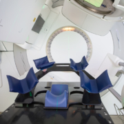
By far the biggest reason why people will arrange cancer care abroad is that we have cutting-edge technology, research and expertise that is unavailable anywhere else in the world.
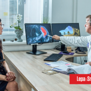
From specialist diagnostics and treatment planning to a wide range of radiotherapy treatments tailored to the type of cancer and your particular needs, many people consider this to seek out a second opinion if their primary option is unavailable.
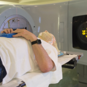
Certain types of treatments, such as stereotactic radiosurgery, are not always available locally and may not be found at any clinic in some countries, making travel essential for high-intensity treatment for certain types of cancer.
Technology aside, if you have a complex or rare condition, you may be referred to an international specialist who can help with a treatment that may otherwise be impossible to treat.
Alternatively, other treatment pathways that are less invasive or more effective may only be available abroad, and whilst there will be an effective course of treatment available at home, one better suited to your overall health goals may only be available internationally.
Lower Waiting Times
Depending on the type of cancer you are diagnosed with, you may be placed on a waiting list for certain types of specialist treatment, which can lead to you waiting months, if not years, to get the care you should have as soon as possible.
Waiting lists and which conditions get priority can vary considerably by country, region or province, depending on access, demand and prognosis.
This can particularly be the case in the UK, where waiting times can vary significantly for certain complex conditions.
Opting for care in Austria can significantly reduce waiting times by avoiding the waiting list. Some doctors will outright suggest doing so or work with you to find an international referral if they believe you need treatment more quickly than it is available domestically.
We have special provisions for international patients, allowing for little waiting time between initial consultation, finalisation of visa documents, arriving in the country and starting treatment.
Alternatively, if you want to pursue a more aggressive form of treatment when you have been suggested active monitoring, seeking treatment abroad can be an alternative option in consultation with your doctor.
Value-driven care
Depending on where you reside and what treatments are available through your public health system, an equivalent treatment may ultimately cost more to stay in your home country than it may to travel and stay abroad.
This is particularly true in countries such as the US, where cancer treatment can be significantly, if not prohibitively expensive if you do not have adequate health insurance or your provider decides not to cover your costs.
Travelling to another country for medical treatment is also a popular option as it can be more cost-effective for people in countries with prohibitive private healthcare costs.
Even in other countries where the disparity between treatment costs is not as extreme, international treatment allows for certain alternative treatment pathways to deliver value-driven care with faster access compared to places like the UK and the US.
Privacy During Treatment
Some people want to be treated in a more secluded environment, either to avoid unwanted attention, to avoid worrying people or to try and mitigate some of the stress that can come with trying to juggle work and public service with cancer care.
Specialist cancer clinics understand how many different factors can affect treatment and will ensure complete confidentiality if this happens to be a priority.