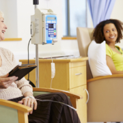
Travelling abroad for treatment is a major commitment to your health, as you get access to state-of-the-art equipment, elite specialists in the field of oncology and access to a wide range of multidisciplinary therapies and treatments not always available in your home country.
We know the importance of the entire care experience when treating cancer, so at our international radiotherapy centre in Vienna, we can provide assistance at every step of your treatment, from advice on securing necessary visas, arranging accommodation and providing continued support throughout your stay.
Part of the considerations will include the travel arrangements, both outbound to Austria and back home. The latter is of particular importance, as depending on the nature of your treatment plan, you may need to spend some time resting and recuperating before you are deemed fit to fly.
However, whether to begin the next stage of your recovery at home or to avoid taking too much time away from work, home and family commitments following a one-day treatment such as some stereotactic radiosurgery procedures, you may be asking when you should return home following the end of treatment.
The short answer is that you should return when your doctor agrees that you are fit to fly and you feel well enough to do so.
To explain why, it is important to understand how treatment may affect travel arrangements, how travel can affect your initial recovery, and what you should do if you are advised to remain in Vienna.
How Is Travel Affected By Cancer Treatment?
Travelling long distances can be stressful on the body, and flying in particular exerts several forces, changes in oxygen levels in the cabin and atmospheric pressures that are typically well-tolerated by the body but can cause issues following certain types of treatment or if you are feeling unwell.
In many cases, this will not affect your ability to fly, but in certain situations, caution may be advised in some cases.
These situations include following conventional surgery (but not stereotactic radiosurgery), if you have a lower red blood cell count, feel breathless or light headed climbing a flight of stairs or are immunocompromised and are at greater risk of catching an infectious disease,
Even if there are no serious risks to travel, if you are feeling particularly fatigued or in pain, a prolonged journey in a confined space could be more uncomfortable than it needs to be.
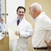
If you are concerned about return travel arrangements, discuss them with your cancer care team. They will be able to provide tailored advice regarding whether it is safe to travel and any precautions you may need to take.
How Does Radiotherapy Affect Travel Arrangements?
Unlike surgery or chemotherapy, which typically require a few weeks to recover before flying, radiotherapy can vary depending on your exact treatment and how you have been progressing with the early stages of your recovery.
In many cases, you can theoretically travel immediately after your final dose of radiotherapy, although for practical reasons such as existing travel itineraries and the time required for your cancer doctor to let the airline know that you are fit to fly and if you require any specific arrangements.
In some cases, you might be particularly sensitive, sore or fatigued after treatment, and so it might be recommended that you wait until you have fully recovered or feel well enough to travel. If you have waves of energy, it may be advised to arrange travel for a day when you feel more ready for it.
Both you and your cancer team will ultimately know when the time is right, and it will be one of several topics that will be discussed during the final treatment appointments.
What Should You Do If You Are Advised To Stay?
We will help and support you every step of the way, from arranging an extension to your accommodation, travel arrangements and follow-up appointments to check in on your health and make sure you are ready to go.
Other than contacting us if you feel your symptoms suddenly improve or get worse, try to enjoy your time in Vienna. Reducing stress is a key step towards your recovery, and taking the time to relax, enjoy the sights and sounds of the historic city and having a little holiday is all part of the process of treatment and recovery.