A diagnosis of cancer can often change one’s relationship with time. The early stages of testing and treatment planning can often be hectic and overwhelming, but there is also a natural desire to start treatment as soon as possible to maximise your options.
There can be delays, either to allow for second opinions or due to the availability of certain types of specialist treatments which could be particularly helpful in your situation.
At that point, there are usually various options available, but one decision a growing number of people are making is to seek medical treatment from specialists abroad.
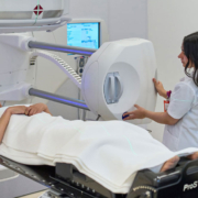
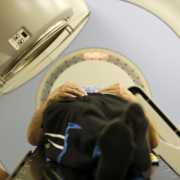
Our international radiotherapy centre is designed to make your medical care in Austria as simple and stress-free as possible. We offer detailed treatment plans, itineraries, help with transport and accommodation, and help you navigate the process of arranging your stay in Vienna.
When is it the best choice for your health? What should you consider before buying any tickets? Is it always an option? And what do you need to know before you fly out for treatment?
Why Do People Consider Medical Treatments Abroad?
Travelling abroad for medical treatment, often defined under the broad banner of “medical tourism”, happens for all sorts of different reasons, even discounting the mental and physical benefits of travelling abroad and seeing the sights in between appointments.
Whilst every patient that we see at our centre has their own story, we have found that many of the reasons for seeking medical care abroad can be grouped into one of three categories.
Second Opinions
One of the biggest reasons for seeking medical care abroad is to access the specialist expertise necessary for an effective second opinion.
In many medical fields, especially oncology, doctors will specialise in specific elements of practice, which means that both their diagnostic and treatment approaches can vary from practitioner to practitioner.
If an initial treatment plan suggests that surgery or a lengthy course of chemotherapy would be the only options, a second opinion may find that a targeted radiotherapy course could be considered as an alternative treatment option in some cases..
In particular, some rare conditions such as rarer types of cancer may require a second opinion from an oncologist with experience treating it.
Faster Care
In some regions, patients may face longer waiting times or have limited access to certain specialist treatments, prompting them to explore treatment options elsewhere.
In some cases, domestic private cancer care can be more expensive than travelling, particularly for other residents of the European Union, and this can make a trip to Austria the best way to access the care you need as soon as possible.
More Options
Not every area has the same access to treatment options, and this can affect the recommendations an oncologist and cancer care team can offer.
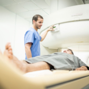
For example, if highly accurate high-dose radiotherapy is unavailable, the team may suggest surgery followed by a broader course of radiotherapy to target any remaining cancer cells.
Having access to a wider range of treatment options may help patients and their medical teams choose the most appropriate treatment approach for their individual circumstances.
What Should You Consider Before Travelling Abroad For Treatment?
As with any other cancer treatment, there is a lot to consider, and it is worth asking your cancer team about any questions you have concerning treatment or the plan for how you will receive it.
Here are some of the most important factors to consider before travelling internationally.
Doctor’s Advice And Procedure
Every country of departure and destination has their own laws and guidance surrounding second opinions and travelling abroad for the purpose of seeking medical treatment, and it is very important to seek advice from your doctor and let them know of your plans.
In some cases, funding can be available, or your treatment plan qualifies under a relevant medical insurance scheme, whilst in other cases you may need to pay the full amount. All of this can be discussed with your potential international clinic.
Is It Safe To Travel?
Depending on the progression of your cancer or the effects of other treatments, travelling to an international clinic may require additional interventions or arrangements following a fitness to fly assessment.
How Will Your Aftercare Be Managed?
Given the schedule of any cancer treatment, it is important to know what your next steps will be following the last appointment, how long you will stay in the country you are receiving treatment and how you will know you are medically cleared and healthy enough to travel back home.